Tissue clearing with high-n reagents is essential for 3D tissue imaging. However, the current liquid-based clearing condition and dye environment suffer from solvent evaporation and photobleaching, causing difficulties in maintaining the tissue optical and fluorescent features
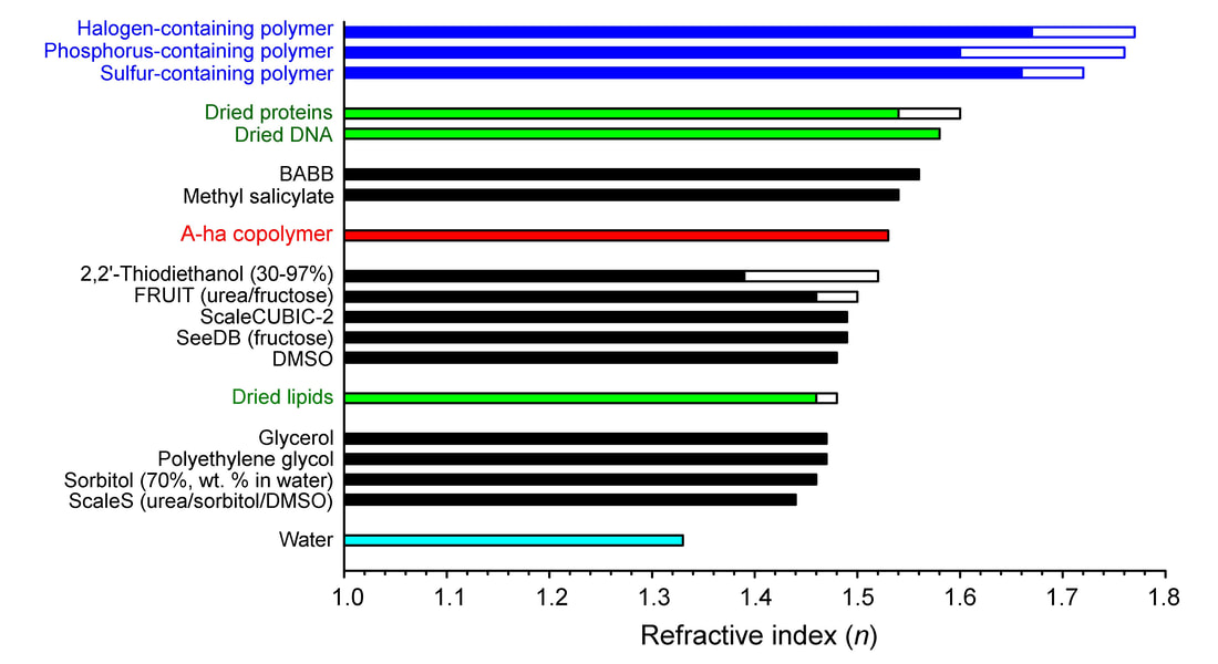
Gladstone-Dale equation: (n-1)/ρ=constant
Increase in n of a material (or reduction in light speed in material) vs. vacuum (n=1) is proportional to the density ρ
In polymer science, the macromolecules formed via covalent bonding are generally denser (higher ρ, and thus higher n) than the monomers in solution prior to polymerization. The bonding reaction (polymerization) thus provides an effective way to increase the density of an organic mixture, creating a high-n environment for optical imaging
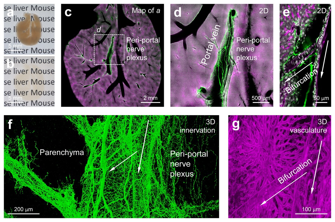
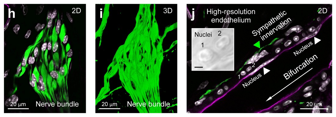
1080pHD video: 3D fluorescence imaging of mouse liver in A-ha copolymer. Still images (left panels) show: i) transparent mouse liver in the A-ha copolymer, ii) fluorescent tissue map of the liver, and iii) hepatic nerves following the portal vein in extension. A magnified and in-depth recording of the perivascular innervation (asterisk) is presented in the right panel (arrow). The overlay of transmitted light and fluorescence signals (00:16-00:54) enhances the presentation. Green, sympathetic nerves (tyrosine hydroxylase+, immunohistochemistry); magenta, blood vessels (perfusion labeling of endothelium); white, nuclei (DAPI, chemical dye staining). All three types of fluorescent labeling are compatible with the A-ha copolymer embedding for high-resolution 3D microscopy.
Antifade imaging in high-n polymer (AF647-conjugated anti-CD31)
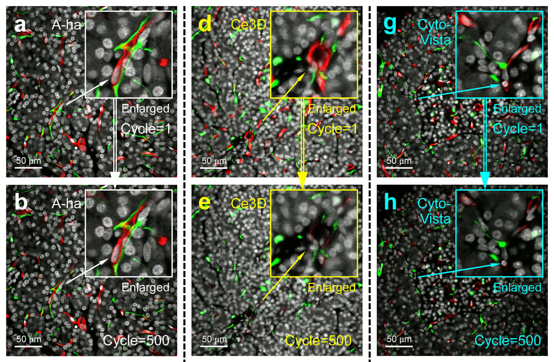
Antifade (time series) test of human pancreas imaging in A-ha copolymer. Tissues were labeled with DAPI (white), anti-S100B (green, standard IHC), and anti-CD31 (red, AF-647-conjugated primary antibody). x40 objective was used to acquire 500 images from the same 320×320-µm region.
1080pHD video: Multimodal imaging of peri-lesional ganglia in human pancreas
Part 1 (00:00-00:12) shows matched H&E and fluorescence images
Part 2 (00:13-00:52) shows matched stereomicroscopic and fluorescence images
Part 1 (00:00-00:12) shows matched H&E and fluorescence images
Part 2 (00:13-00:52) shows matched stereomicroscopic and fluorescence images
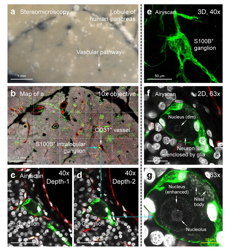
Magnified image & 1080pHD video: 3D/Airyscan super-resolution imaging of human intrapancreatic ganglion in high-n polymer
1080pHD video gallery of PGP9.5-labeled human intrapancreatic ganglia
|
|
|
|
|
|